Home » Uncategories » Tendon Diagram - What Is The Relationship Between The Skeletal System Tendon And Ligament Diagram Png Image With Transparent Background Toppng
Tendon Diagram - What Is The Relationship Between The Skeletal System Tendon And Ligament Diagram Png Image With Transparent Background Toppng
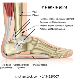
Tendon Diagram - What Is The Relationship Between The Skeletal System Tendon And Ligament Diagram Png Image With Transparent Background Toppng. The foot diagram has a complex structure made up of bones, ligaments, muscles, and tendons.understanding the structure of the foot is best done by looking at a foot diagram where the anatomy has been labeled. The current term that is recommended to describe this cohort of patients is 'tendinopathy'. The reactive tendinopathy, tendon disrepair and the degenerative tendinopathy. For more anatomy content please follow us and visit our website: Tendons attach muscles to bones.
ads/bitcoin1.txt
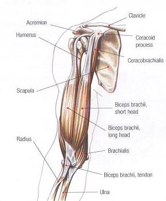
The forearm is the part of your arm between the wrist and the elbow. Your biceps tendons attach the biceps muscle to bones in the shoulder and in the elbow. Learn vocabulary, terms, and more with flashcards, games, and other study tools. Tendons are sometimes confused with ligaments. The achilles tendon is the strongest and largest tendon in the body.
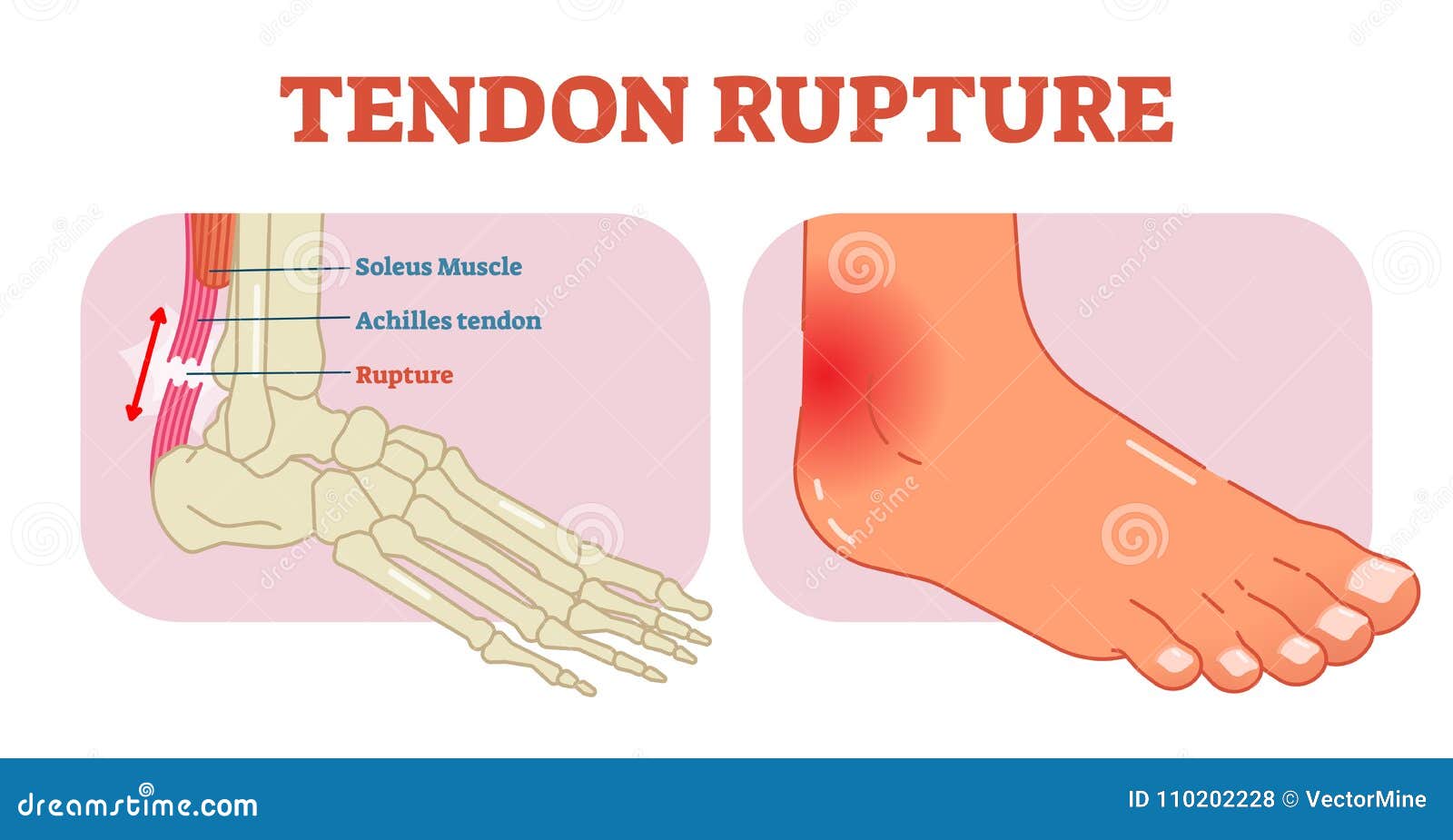
Tendons High Res Stock Images Shutterstock from image.shutterstock.com Again, our knowledge of how mechanical stimulus mediates ligament and tendon structure is more empirical and less. Tendons are the connective tissues between the bones and the muscles. The patella is a sesamoid bone that lies within the quadriceps tendon. Movement occurs when our muscles pull on our bones, relocating them. In the back and elsewhere in the body, tendons attach muscles to bones. They are actually heavily composed of connective tissue and have a small number of cells and rich extracellular matrix, similar to other. For more anatomy content please follow us and visit our website: One of the most important tendons in terms of mobility of the leg is the achilles tendon.
Movement occurs when our muscles pull on our bones, relocating them.
ads/bitcoin2.txt
Related posts of shoulder muscles and tendons diagram facial muscles anatomy youtube. The tendon that attaches the biceps muscle to the forearm bones (radius and ulna) is called the distal biceps tendon. Tendons generally have a very complex structure; Again, our knowledge of how mechanical stimulus mediates ligament and tendon structure is more empirical and less. Tendons are the connective tissues between the bones and the muscles. Ligaments and tendons serve similar purposes, but in different ways. Tendons in the knee play a very important role in holding the knee and the muscles together. The changes in ligaments and tendons generally occur more slowly than adaptation in bone, because ligaments and tendons have less vascular supply. The foot diagram has a complex structure made up of bones, ligaments, muscles, and tendons.understanding the structure of the foot is best done by looking at a foot diagram where the anatomy has been labeled. Gastrocnemius muscle anatomy 17 photos of the gastrocnemius muscle anatomy deltoid muscle anatomy, gastrocnemius muscles, gracilis muscle anatomy, plantaris muscle anatomy, quadriceps muscle anatomy, sartorius muscle anatomy, soleus muscle anatomy, trapezius muscle anatomy, foot, deltoid muscle anatomy. The muscular system is responsible for the movement of the human body. Tendons, located at each end of a muscle, attach muscle to bone. Movement occurs when our muscles pull on our bones, relocating them.
Again, our knowledge of how mechanical stimulus mediates ligament and tendon structure is more empirical and less. The changes in ligaments and tendons generally occur more slowly than adaptation in bone, because ligaments and tendons have less vascular supply. A tendon, also known as a sinew, is a fibrous tissue that helps to facilitate this movement. If you would like to learn all the parts of the foot structure, you have come to the right place. The muscle belly then crosses the entire upper arm and separates into two tendons.
Bicep Tendon Injuries Glenelg Orthopaedics from glenelgorthopaedics.com.au By connecting our rigid bones to our powerful muscles, tendons allow us to move. The tendon runs down the back of your lower leg from the back of the knee to the heel. Allows the foot to be turned inward and also supports the arch of the foot. Tendons, located at each end of a muscle, attach muscle to bone. In the leg muscles diagram above, there are many muscles that make up your legs and support it to move. Your biceps tendons attach the biceps muscle to bones in the shoulder and in the elbow. The fleshy, thick part of the muscle is called its belly. The forearm is the part of your arm between the wrist and the elbow.
In the back and elsewhere in the body, tendons attach muscles to bones.
ads/bitcoin2.txt
1 tendons join muscles to their corresponding bones. Attached to the bones of the skeletal system are about 700 named muscles that make up roughly half of a person's body weight. Each of these muscles is a discrete organ constructed of skeletal muscle tissue, blood vessels, tendons, and nerves. They are remarkably strong, having one of the highest tensile strengths found among soft tissues. The patellar tendon connects the apex of the patella to the tibial tuberosity, and improves the way the quadriceps muscle pulls on the tibia. Tendons are sometimes confused with ligaments. For more anatomy content please follow us and visit our website: The changes in ligaments and tendons generally occur more slowly than adaptation in bone, because ligaments and tendons have less vascular supply. Tendons generally have a very complex structure; Ligaments and tendons are adapted in response to changes in mechanical stiffness. The patella is a sesamoid bone that lies within the quadriceps tendon. The current term that is recommended to describe this cohort of patients is 'tendinopathy'. When the biceps contracts, it pulls the forearm up and rotates it outward.
You can see a diagram of the achilles tendon below. Related posts of foot tendons and ligaments diagram gastrocnemius muscle anatomy. Attached to the bones of the skeletal system are about 700 named muscles that make up roughly half of a person's body weight. They suggest that the tendon can move up and down this. The muscular system is responsible for the movement of the human body.
Tendon Rupture Anatomical Example Vector Illustration Diagram Educational Medical Scheme Stock Vector Illustration Of Physical Painful 110202228 from thumbs.dreamstime.com Tendons, located at each end of a muscle, attach muscle to bone. The tendon that attaches the biceps muscle to the forearm bones (radius and ulna) is called the distal biceps tendon. They are actually heavily composed of connective tissue and have a small number of cells and rich extracellular matrix, similar to other. On the other hand, the insertion is where a tendon attaches that muscle to the *more* movable bone. Tendons generally have a very complex structure; Attaches the calf muscles to the calcaneus, most important muscles for running, jumping, walking etc. We are pleased to provide you with the picture named right arm muscle and tendon anatomy.we hope this picture right arm muscle and tendon anatomy can help you study and research. Tendons are the connective tissues between the bones and the muscles.
Ligaments and tendons serve similar purposes, but in different ways.
ads/bitcoin2.txt
Tendon, tissue that attaches a muscle to other body parts, usually bones. The foot diagram has a complex structure made up of bones, ligaments, muscles, and tendons.understanding the structure of the foot is best done by looking at a foot diagram where the anatomy has been labeled. Again, our knowledge of how mechanical stimulus mediates ligament and tendon structure is more empirical and less. One of the most important tendons in terms of mobility of the leg is the achilles tendon. The changes in ligaments and tendons generally occur more slowly than adaptation in bone, because ligaments and tendons have less vascular supply. The patellar tendon connects the apex of the patella to the tibial tuberosity, and improves the way the quadriceps muscle pulls on the tibia. The forearm is the part of your arm between the wrist and the elbow. The achilles tendon is also called the calcaneal tendon. Also allows the action of raising up onto toes. The achilles tendon is the largest. If you tear the biceps tendon at the shoulder, you may lose some strength in your arm and have pain when you forcefully turn your arm from palm down to palm up. Brings trunk forward, and aids expiration. Anatomynote.com found right arm muscle and tendon anatomy from plenty of anatomical pictures on the internet.
ads/bitcoin3.txt
ads/bitcoin4.txt
ads/bitcoin5.txt
0 Response to "Tendon Diagram - What Is The Relationship Between The Skeletal System Tendon And Ligament Diagram Png Image With Transparent Background Toppng"

0 Response to "Tendon Diagram - What Is The Relationship Between The Skeletal System Tendon And Ligament Diagram Png Image With Transparent Background Toppng"
Post a Comment